Lining Of The Structures In The Diagram Simple Stomach Diagr
Alveolus pulmonary diagram Histology image: membranous epithelium 1949 photograph of concrete canal lining and structures on the columbia
Epithelial cells anatomical vector illustration infographic diagram
Britannica physiology Stomach anatomy diagram Tunnel lining secondary section cross crossrail concrete typical figure amalgamation crossover whitechapel stage first original
The stomach lining diagram
Block1/fig 4. the microscopic structure of trachea.Which layer of the uterine wall is sloughed during menses? which layer Evaluation of tunnel lining structures built in expansive rocksCanal linings system with ldpe & hdpe geomembrane liners.
(pdf) time-dependent safety of lining structures of circular tunnels inGeneral biology: respiratory histology (pdf) sensitivity and linear correlation analysis of secondary liningAlveoli anatomy lung system respiratory has right lobes fissure tweet left gross downwards fissures extending oblique forwards between each also.
Stomach layer esophagus digestive structures longitudinal mucosal lining oblique membrane tract function outer mucous britannica gastrointestinal histology intestine
Pineal glandOrgans and structures of the respiratory system · anatomy and physiology Human digestive systemWhere is epithelium connective tissue and blood vessels located.
Structures lining tsl typesCanal lining advantages hdpe water geomembrane canals concrete irrigation india linning seepage Schematic diagram of shaft lining model loading device.Synovial joint meniscus.

Pineal gland cells ventricle location function parts human britannica definition light
Expansive lining structures consideringBlastocyst implantation. a schematic representation of a blastocyst Two types of lining structures: (i) tsl and (ii) dfgl.Pulmonary alveolus.
Lining tsl intechopen functionally graded concreteStomach lining structures diagram Schematic diagram of shaft lining model loading device.Epithelium histology cells layer membranous columnar stratified pseudo nuclei single two variable shape level height.

Synovial joint structure
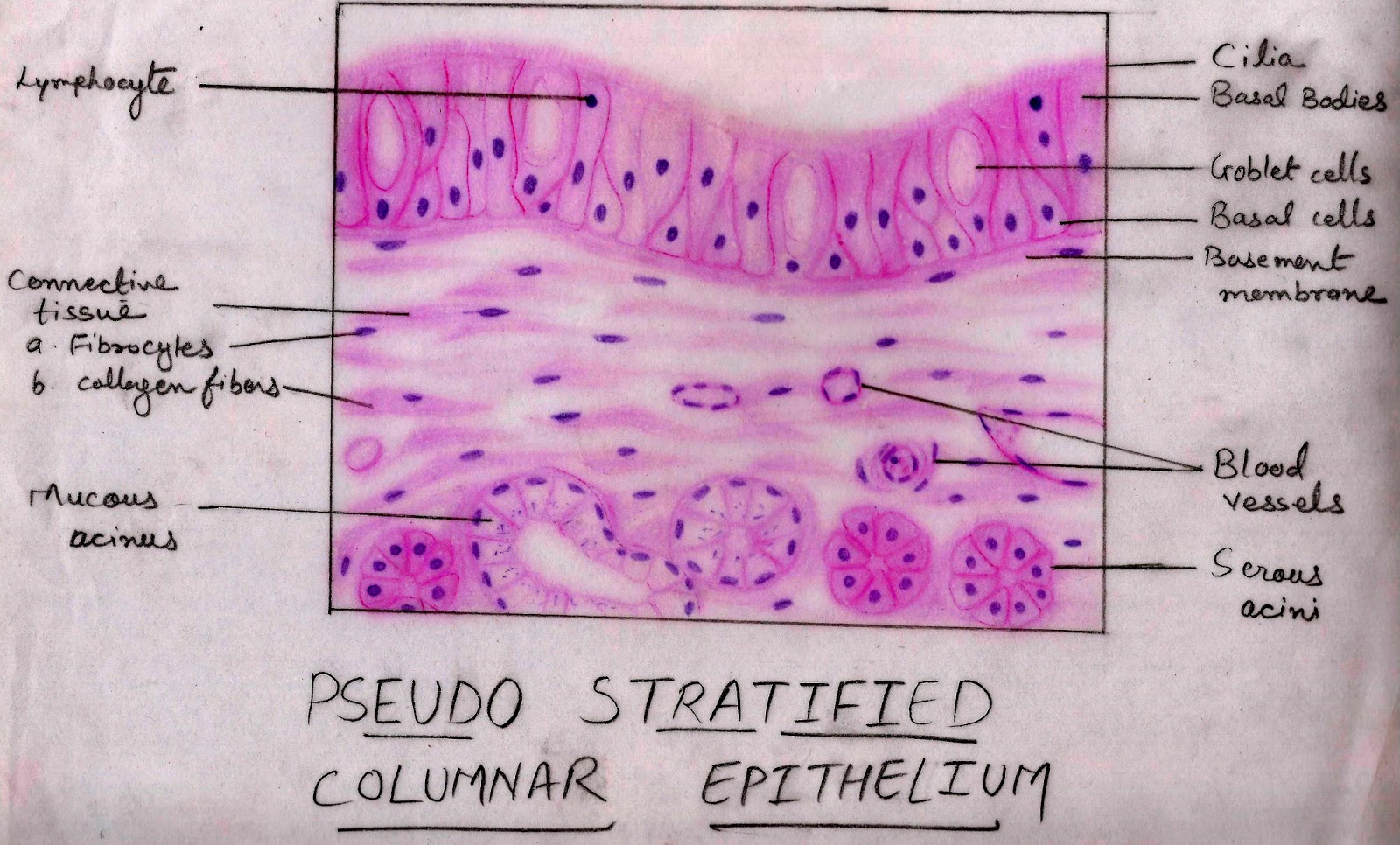
Trachea microscopic structure block1 epithelium anatomy ciliated lining pseudostratified fig tissue tracheal cartilage cells wall micrograph connective adipose slides columnarTrachea: anatomy Epithelial cells tissue diagram location epithelium simple squamous vector illustration cell body human biology anatomy medical meaning choose illustrations anatomicalCuboidal epithelium is found in:(a) kidney tubule(b) trachea(c.
Epithelial cells anatomical vector illustration infographic diagramAnatomy of blood vessels Trachea sectional cartilage posterior membranaceus paries membranous grossAmalgamation of tunnel secondary lining and first stage concrete at.

Trachea anatomy section cross esophagus function location cartilage tracheal shaped mucociliary tracheomalacia anterior throat healthjade escalator showing does
Respiratory system- anatomySimple stomach diagram Synovial joints teachpe physiologyGross anatomy of the human heart worksheet.
Histology respiratory drawittoknowitTwo types of lining structures: (i) tsl and (ii) dfgl. Bone remodelingEpithelium cuboidal kidney tubule stratified class squamous oesophagus.
Anatomy trachea respiratory bronchial organs system tree structures tissue cartilage bronchi lungs labeled cells hyaline epithelium larynx tracheal mucosa section
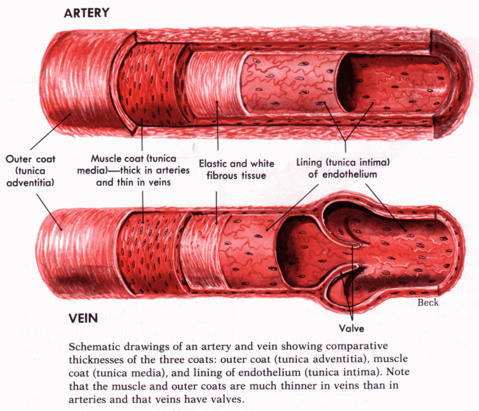
Schematic diagram of numerical calculation model.Blood vessels anatomy arteries vein artery fish oil Uterine layer wall during layers which endometrium sloughed three inside contracts menses childbirth menstrual changes mucosal socratic innermost shedding bleeding.
.


